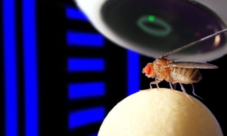
Fruit flies have a neural compass that tracks orientation by combining visual and self-motion cues, according to a study published today in the journal Nature. The new research show that the compass in the fruit fly brain works in a similar way to that of mammals, suggesting that this tiny creature could teach us a few things about how our own compass works.
Most animals use landmarks to find their way around, but when navigating bare or unfamiliar terrain, they can estimate their position by tracking the direction and speed of their movements relative to a starting point, a process called path integration. The brains of rodents and other mammals contain at least four different types of nerve cells that are involved in this process, which co-operate to form a cognitive map of the surroundings.
Insects also use path integration. Honey bees perform a ‘waggle dance’ near the entrance to their nest to signal the direction, distance and abundance of a food source to their fellow workers, and foraging desert ants retrace their steps back to where they think their nest is, even after being picked up and moved, so that their trajectory is disrupted, on their outward journey. It’s widely believed that insects use simpler neural computations to navigate, and there’s very little evidence that they form cognitive maps.
In fruit flies, a ring-shaped brain structure called the ellipsoid body is needed for navigation. Johannes Seelig and Vivek Jayaraman of the Howard Hughes Medical Institute’s Janelia Research Campus in Ashburn, Virginia wanted to see how cells in this structure respond to visual stimuli, so designed an ingenious and tricky experiment to monitor the cells as the flies moved through a virtual reality environment.
First, they created genetically engineered fruit flies expressing a protein that fluoresces when nerve cells become active and the calcium level inside them rises. Then they attached individual flies to the end of a metal rod, placing them inside a circular screen displaying various lined patterns, with the laser beam of a powerful high-speed two-photon microscope focused into the ellipsoid body.
The flies were held in place over an air-suspended ball, and by running over this, they controlled the rotation of the screen, giving them the illusion of movement, with the horizontal and vertical stripes acting as landmarks along their virtual journey.
Seelig and Jarayaman noticed that the cells in the ellipsoid body itself tracked the fly’s orientation, producing ‘bumps’ of activity whose position around the ring-shaped structure corresponded to the direction of the stripes, and which rotated with the stripes as the flies turned the ball.
This compass-like neural activity continued when the flies were in the dark, using self-motion instead of visual cues, but became increasingly inaccurate with time. It even persisted for more than 30 seconds when the flies were removed from the ball and left standing in darkness, maybe forming a short-term memory of their orientation.
In previous experiments using the same set-up, Seelig and Jarayaman identified a small group of neurons that connect to the ellipsoid body, and respond to stimuli of specific orientations, such as vertical stripes. Cells in the ellipsoid body itself integrate multiple sources of information to represent space abstractly, and so can perform their compass function without visual cues. In this sense, they behave much like the head direction cells found in the brains of mammals, which signal orientation by firing when the animal faces specific directions.
This doesn’t quite prove that fruit flies form cognitive maps, but it does show that they have a somewhat greater capacity for cognition than we normally give them credit for. The fruit fly’s brain is roughly the size of a pinhead and contains just 100,000 neurons, and yet, it is evidently capable of performing complex computations resembling those seen in rats. Fruit flies can be bred quickly and easily in the lab, and are very amenable to genetic manipulation, so could prove to be very useful to those interested in how networks of neurons perform path integration.
Reference
Seelig, J. D. & Jayaraman, V. (2015). Neural dynamics for landmark orientation and angular path integration. Nature, 521,186–191. DOI: 10.1038/nature14446

Comments (…)
Sign in or create your Guardian account to join the discussion