Summary: Study confirms the role the corpus callosum plays in language lateralization.
Source: HSE
A study by the HSE Centre for Language and Brain has confirmed the role of the corpus callosum in language lateralization, the distribution of language processing functions between the brain’s hemispheres.
The authors came up with an innovative language task for their study subjects and applied advanced neuroimaging methods to the data collected.
A paper on their findings has been published in PLoS ONE.
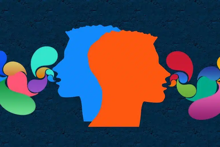
Functional asymmetry between the two cerebral hemispheres in performing higher-level cognitive functions is a major characteristic of the human brain. For example, the left hemisphere plays a leading role in language processing in most people. However, between 10% and 15% of the human population also use the right hemisphere to varying degrees for the same task.
Traditionally, language lateralization to the right hemisphere was explained by handedness, as it is mainly found in left-handed and ambidextrous (using both hands equally well) individuals. But recent research has demonstrated a genetic difference in the way language is processed by left-handed and ambidextrous people.
In addition to this, some right-handed people also involve their right hemisphere in language functions.
These findings prompted the scientists to consider alternative explanations—in particular, by looking at brain anatomy to find out why language functions can shift to the right hemisphere.
Researchers at the HSE Centre for Language and Brain hypothesized that language lateralization may have something to do with the anatomy of the corpus callosum, the largest commissural tract in the human brain connecting the two cerebral hemispheres.
The researchers asked 50 study participants to perform a sentence completion task. The subjects were instructed to read aloud a visually presented Russian sentence and to complete it with an appropriate final word (e.g., “Teper ministr podpisyvaet vazhnoe…”—”Now the minister is signing an important …”).
At the same time, the participants’ brain activity was recorded using functional magnetic resonance imaging (fMRI). Additionally, the volume of the corpus callosum was measured in each subject.
A comparison between the fMRI data and the corpus callosum measurements revealed that the larger the latter’s volume, the less lateralization of the language function to the right hemisphere was observed.
It can be said that in processing language, the brain tends to use the left hemisphere’s resources efficiently and to suppress, by means of the corpus callosum, any additional involvement of the right hemisphere. The larger a person’s corpus callosum, the less involved their right hemisphere is in language processing (and vice versa).
This finding is consistent with the inhibitory model suggesting that the corpus callosum inhibits the action of one hemisphere while the other is engaged in cognitive tasks.
“The study’s innovative design and use of advanced neuroimaging have made this conclusion possible.
“Brain lateralization in language processing is usually hard to measure accurately, as typical speech tasks used in earlier studies (eg image naming, selecting words that begin with a certain letter or listening to speech) tend to cause activation only in some parts of the brain responsible for language functions but not in others.
“Instead, we developed a unique speech task for fMRI—sentence completion—which reliably activates all language areas of the brain,” says Olga Dragoy, Director of the HSE Centre for Language and Brain.
It is important to add that the authors reconstructed the volume and properties of the corpus callosum from MRI data using an advanced tractography technique: constrained spherical deconvolution (CSD).
This is more suitable than traditional diffusion tensor imaging for modeling crossing fibers in the smallest unit of volume, the voxel (3D pixel), and is therefore more reliable.
About this language and neuroscience research news
Author: Anastasia Lobanova
Source: HSE
Contact: Anastasia Lobanova – HSE
Image: The image is in the public domain
Original Research: Open access.
“Greater volumes of a callosal sub-region terminating in posterior language-related areas predict a stronger degree of language lateralization: A tractography study” by Victor Karpychev et al. PLOS ONE
Abstract
Greater volumes of a callosal sub-region terminating in posterior language-related areas predict a stronger degree of language lateralization: A tractography study
Language lateralization is the most intriguing trait of functional asymmetry for cognitive functions.
Nowadays, ontogenetic determinants of this trait are largely unknown, but there are efforts to find its anatomical correlates. In particular, a white matter interhemispheric connection–the corpus callosum–has been proposed as such.
In the present study, we aimed to find the association between the degree of language lateralization and metrics of the callosal sub-regions.
We applied a sentence completion fMRI task to measure the degree of language lateralization in a group of healthy participants balanced for handedness.
We obtained the volumes and microstructural properties of callosal sub-regions with two tractography techniques, diffusion tensor imaging (DTI) and constrained spherical deconvolution (CSD).
The analysis of DTI-based metrics did not reveal any significant associations with language lateralization. In contrast, CSD-based analysis revealed that the volumes of a callosal sub-region terminating in the core posterior language-related areas predict a stronger degree of language lateralization.
This finding supports the specific inhibitory model implemented through the callosal fibers projecting into the core posterior language-related areas in the degree of language lateralization, with no relevant contribution of other callosal sub-regions.