saccular aneurysm
Also found in: Dictionary, Thesaurus, Encyclopedia.
aneurysm
[an´u-rizm]a sac formed by the localized dilatation of the wall of an artery, a vein, or the heart.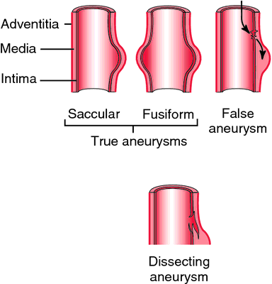 adj., adj aneurys´mal. The chief signs of an arterial aneurysm are the formation of a pulsating tumor, and often a bruit (aneurysmal bruit) heard over the swelling. Sometimes there are symptoms from pressure on contiguous parts.
The most common site for an arterial aneurysm is the abdominal aorta. A true aneurysm results from formation of a sac by the arterial wall with at least one unbroken layer. It is most often associated with atherosclerosis. A false aneurysm usually is caused by trauma. In this case, the wall of the blood vessel is ruptured and blood escapes into surrounding tissues and forms a clot. Because of pressure within the clot arising from the heart's contractions, the clot often pulsates against the examiner's hand as does a true aneurysm.
Although atherosclerosis is responsible for most arterial aneurysms, any injury to the middle or muscular layer of the arterial wall (tunica media) can predispose the vessel to stretching of the inner and outer layers of the artery and the formation of a sac. Other diseases that can lead to an aneurysm include syphilis, cystic medionecrosis, certain nonspecific inflammations, and congenital defects in the artery.
It is possible for a person to be unaware of a small aneurysm for years. About 80 per cent of all abdominal aneurysms are palpable and may be noticed on a routine physical examination. One should be particularly alert to the possibility of an aneurysm in persons with a history of cardiovascular disease, hypertension, or peripheral vascular disease.
Aneurysms tend to increase in size, presenting a problem of increasing pressure against adjacent tissues and organs and a danger of rupture. When an aneurysm ruptures, a critical situation ensues. The patient with a ruptured aortic aneurysm exhibits severe pain and blood loss, leading to shock. A ruptured cerebral aneurysm produces neurologic symptoms and can resemble the clinical picture of stroke syndrome.
Treatment of aneurysm depends on the vessel involved, size of the aneurysm, and general health status of the patient.

Classification of aneurysms. All three tunica layers are involved in true aneurysms (fusiform and saccular). In false aneurysms, blood escapes between tunica layers and they separate. If the separation continues, a clot may form, resulting in a dissecting aneurysm. From Copstead and Banasik, 2000.
The most common site for an arterial aneurysm is the abdominal aorta. A true aneurysm results from formation of a sac by the arterial wall with at least one unbroken layer. It is most often associated with atherosclerosis. A false aneurysm usually is caused by trauma. In this case, the wall of the blood vessel is ruptured and blood escapes into surrounding tissues and forms a clot. Because of pressure within the clot arising from the heart's contractions, the clot often pulsates against the examiner's hand as does a true aneurysm.
Although atherosclerosis is responsible for most arterial aneurysms, any injury to the middle or muscular layer of the arterial wall (tunica media) can predispose the vessel to stretching of the inner and outer layers of the artery and the formation of a sac. Other diseases that can lead to an aneurysm include syphilis, cystic medionecrosis, certain nonspecific inflammations, and congenital defects in the artery.
It is possible for a person to be unaware of a small aneurysm for years. About 80 per cent of all abdominal aneurysms are palpable and may be noticed on a routine physical examination. One should be particularly alert to the possibility of an aneurysm in persons with a history of cardiovascular disease, hypertension, or peripheral vascular disease.
Aneurysms tend to increase in size, presenting a problem of increasing pressure against adjacent tissues and organs and a danger of rupture. When an aneurysm ruptures, a critical situation ensues. The patient with a ruptured aortic aneurysm exhibits severe pain and blood loss, leading to shock. A ruptured cerebral aneurysm produces neurologic symptoms and can resemble the clinical picture of stroke syndrome.
Treatment of aneurysm depends on the vessel involved, size of the aneurysm, and general health status of the patient.
arteriosclerotic aneurysm an aneurysm arising in a large artery, most commonly the abdominal aorta, as a result of weakening of the wall in severe atherosclerosis; called also atherosclerotic aneurysm.
arteriovenous aneurysm an abnormal communication between an artery and a vein in which the blood flows directly into a neighboring vein or is carried into the vein by a connecting sac.
atherosclerotic aneurysm arteriosclerotic aneurysm.
bacterial aneurysm an infected aneurysm caused by bacteria.
berry aneurysm (brain aneurysm) a small saccular aneurysm of a cerebral artery, usually at the junction of vessels in the circle of Willis; such aneurysms frequently rupture, causing subarachnoid hemorrhage. Called also cerebral aneurysm.
cardiac aneurysm thinning and dilatation of a portion of the wall of the left ventricle, usually a consequence of myocardial infarction.
cerebral aneurysm berry aneurysm.
cirsoid aneurysm dilatation and tortuous lengthening of part of an artery; called also racemose aneurysm.
compound aneurysm one in which some of the layers of the wall of the vessel are ruptured and some merely dilated; called also mixed aneurysm.
dissecting aneurysm one resulting from hemorrhage that causes lengthwise splitting of the arterial wall, producing a tear in the inner wall (intima) and establishing communication with the lumen of the vessel. It usually affects the thoracic aorta (see aortic dissection) but can also occur in other large arteries. See illustration.
false aneurysm
one in which the entire wall is injured and the blood is contained by the surrounding tissues, with eventual formation of a sac communicating with the artery (or heart). See illustration.
fusiform aneurysm a spindle-shaped aneurysm; see illustration.
infected aneurysm one produced by growth of microorganisms (bacteria or fungi) in the vessel wall, or infection arising within a preexisting arteriosclerotic aneurysm.
mixed aneurysm compound aneurysm.
mycotic aneurysm an infected aneurysm caused by fungi.
racemose aneurysm cirsoid aneurysm.
saccular aneurysm (sacculated aneurysm) a saclike aneurysm; see illustration.
spurious aneurysm
false aneurysm (def. 1).
varicose aneurysm one formed by rupture of an aneurysm into a vein.
Miller-Keane Encyclopedia and Dictionary of Medicine, Nursing, and Allied Health, Seventh Edition. © 2003 by Saunders, an imprint of Elsevier, Inc. All rights reserved.
sac·cu·lar an·eu·rysm
, sacculated aneurysma saclike bulging on one side of an artery.
Synonym(s): ampullary aneurysm
Farlex Partner Medical Dictionary © Farlex 2012
saccular aneurysm
An eccentric aneurysm due to weakening of one side of a vessel wallMcGraw-Hill Concise Dictionary of Modern Medicine. © 2002 by The McGraw-Hill Companies, Inc.
sac·cu·lar an·eu·rysm
, sacculated aneurysm (sak'yū-lăr an'yūr-izm, sak'yū-lā'tĕd)A saclike bulging on one side of an artery.
Medical Dictionary for the Health Professions and Nursing © Farlex 2012
saccular aneurysm
A sac-like ballooning in the wall of an artery that communicates with the vessel by a relatively small opening.Collins Dictionary of Medicine © Robert M. Youngson 2004, 2005
Saccular aneurysm
A type of aneurysm that resembles a small sack of blood attached to the outer surface of a blood vessel by a thin neck.
Mentioned in: Arteriovenous Malformations
Gale Encyclopedia of Medicine. Copyright 2008 The Gale Group, Inc. All rights reserved.