cholangiography
Also found in: Dictionary, Thesaurus, Encyclopedia, Wikipedia.
Related to cholangiography: cholangiogram, cholecystography, Intravenous Cholangiography, operative cholangiography, Percutaneous transhepatic cholangiography, T tube cholangiography
cholangiography
[ko-lan″je-og´rah-fe]x-ray examination of the bile ducts, using a radiopaque dye as a contrast medium. In the intravenous method, the dye is administered intravenously and is excreted by the liver into the bile ducts. X-ray films are taken at 10-minute intervals as the dye is excreted via the cystic, hepatic, and common bile ducts into the intestinal tract. The excretion is usually completed within 4 hours. Preparation of the patient for the intravenous method requires restriction of fluids to concentrate the dye and may also include cleansing of the intestinal tract on the day prior to the examination with a laxative or enema so that fecal material and gas will not obscure the biliary tract.
Sometimes cholangiography is done after surgery of the gallbladder and biliary tract. In this method the radiopaque dye is injected directly into a tube that has been left in the biliary tract since the time of surgery. Films are taken immediately after the dye is injected. If no obstruction is present, the biliary structures fill readily and rapidly empty into the intestinal tract.
When it is necessary for the surgeon to locate gallstones or other obstructive conditions at the time that surgery is being performed, the dye may be injected directly into the bile ducts. Films are taken in the operating room, and obstructions not otherwise discernible can be located and corrected while the patient is still anesthetized.
A patient who is jaundiced cannot undergo either intravenous cholangiography or oral cholecystography. An alternative route for the injection of the contrast dye and visualization of the biliary system is percutaneous transhepatic cholangiography. Under fluoroscopic control, a needle is introduced through the skin and into the liver where the contrast material is deposited. Obstructed and distended bile ducts can then be visualized. After visualization the ducts can be drained via the needle.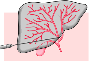

Percutaneous transhepatic cholangiography. The aspirating needle is passed through the patient's skin and liver tissue until the tip penetrates one of the hepatic ducts. Radiopaque medium is then instilled into the biliary tree to enhance radiographic visualization. From Malarkey and McMorrow, 2000.
fine needle transhepatic cholangiography (FNTC) transhepatic cholangiography performed by means of a very fine, highly flexible steel needle (skinny needle).
percutaneous transhepatic cholangiography see cholangiography.
transhepatic cholangiography cholangiography after introduction of radiopaque medium into the biliary system by percutaneous puncture of a bile duct.
transjugular cholangiography cholangiography after catheterization of a hepatic vein via the internal jugular vein in the neck and entry into a bile duct by percutaneous puncture across the wall of the hepatic vein.
Miller-Keane Encyclopedia and Dictionary of Medicine, Nursing, and Allied Health, Seventh Edition. © 2003 by Saunders, an imprint of Elsevier, Inc. All rights reserved.
chol·an·gi·og·ra·phy
(kō-lan'jē-og'ră-fē),Radiographic examination of the bile ducts with contrast medium.
[chol- + G. angeion, vessel, + graphō, to write]
Farlex Partner Medical Dictionary © Farlex 2012
cholangiography
(kō-lăn′jē-ŏg′rə-fē)n.
X-ray examination of the bile ducts after the administration of a radiopaque contrast medium.
cho·lan′gi·o·graph′ic (-ə-grăf′ĭk) adj.
The American Heritage® Medical Dictionary Copyright © 2007, 2004 by Houghton Mifflin Company. Published by Houghton Mifflin Company. All rights reserved.
cholangiography
Imaging Oral, IV or percutaneous administration of radiocontrast excreted into the bile tract, to detect gallstones or visualize bile ducts. See Percutaneous transhepatic cholangiography.McGraw-Hill Concise Dictionary of Modern Medicine. © 2002 by The McGraw-Hill Companies, Inc.
chol·an·gi·og·ra·phy
(kō-lan'jē-og'ră-fē)Radiographic examination of the bile ducts using a contrast medium.
[chol- + G. angeion, vessel, + graphō, to write]
Medical Dictionary for the Health Professions and Nursing © Farlex 2012
cholangiography
X-ray or other imaging examination of the bile ducts, usually after a fluid substance opaque to radiation has been introduced. The main object of cholangiography is to show stones in the bile ducts and gall bladder. The method may be done by direct injection through the skin into the liver (percutaneous, transhepatic cholangiography) or, by way of a flexible endoscope, through the bile duct opening in the duodenum (endoscopic retrograde cholangiography).Collins Dictionary of Medicine © Robert M. Youngson 2004, 2005
Cholangiography
Radiographic examination of the bile ducts after injection with a special dye.
Mentioned in: Bile Duct Cancer, Gallbladder Cancer
Gale Encyclopedia of Medicine. Copyright 2008 The Gale Group, Inc. All rights reserved.