aorta

(redirected from aortal)
Also found in: Dictionary, Thesaurus, Encyclopedia.

aorta

 [a-or´tah] (pl. aor´tae, aortas) (L.)
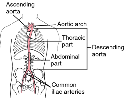
the great artery arising from the left ventricle, being the main trunk from which the systemic arterial system proceeds. It has four divisions: the ascending aorta, the aortic arch, the thoracic aorta, and the abdominal aorta. See Appendix of Arteries and see circulatory system.
 Aorta, arising from the left ventricle, ascending, arching, then descending through the thorax to the abdomen, where it divides into the common iliac arteries. From Dorland's, 2000.
overriding aorta a congenital anomaly occurring in tetralogy of Fallot, in which the aorta is displaced to the right so that it appears to arise from both ventricles and straddles the ventricular septal defect.
Miller-Keane Encyclopedia and Dictionary of Medicine, Nursing, and Allied Health, Seventh Edition. © 2003 by Saunders, an imprint of Elsevier, Inc. All rights reserved.

a·or·ta

, gen. and pl.

a·or·tae

(ā-ōr'tă, ā-ōr'tē), [TA]
A large artery of the elastic type that is the main trunk of the systemic arterial system, arising from the base of the left ventricle and ending at the left side of the body of the fourth lumbar vertebra by dividing to form the right and left common iliac arteries. The aorta is subdivided into: ascending aorta; aortic arch; and descending aorta, which is in turn, divided into the thoracic aorta and the abdominal aorta.
Synonym(s): arteria aorta
[Mod. L. fr. G. aortē, from aeirō, to lift up]
Farlex Partner Medical Dictionary © Farlex 2012

aorta

(ā-ôr′tə)
n. pl. aor·tas or aor·tae (-tē)
The main trunk of the systemic arteries, carrying blood from the left side of the heart to the arteries of all limbs and organs except the lungs.

a·or′tal, a·or′tic adj.
The American Heritage® Medical Dictionary Copyright © 2007, 2004 by Houghton Mifflin Company. Published by Houghton Mifflin Company. All rights reserved.

a·or·ta

, pl. aortae (ā-ōr'tă, -tē) [TA]
A large artery that is the main trunk of the systemic arterial system, arising from the left ventricle and ending at the left side of the body of the fourth lumbar vertebra by dividing to form the right and left common iliac arteries. The aorta is made up of the ascending aorta, aortic arch, and descending aorta, which is divided into the thoracic aorta and the abdominal aorta.
[Mod. L. fr. G. aortē, from aeirō, to lift up]
Medical Dictionary for the Health Professions and Nursing © Farlex 2012

aorta

(ā-ort′ă ) (ā-ort′ē) plural.aortaeaortas [L. aorta fr Gr. aortē, the large artery]
Enlarge picture
MAIN PARTS OF AORTA
Enlarge picture
MAIN PARTS OF AORTA
The main trunk of the arterial system of the body. aortic (ā-or′tĭk), adjective

The aorta is about 3 cm in diameter at its origin in the upper surface of the left ventricle. It passes upward as the ascending aorta, turns backward and to the left (arch of the aorta) at about the level of the fourth thoracic vertebra, and then passes downward as the thoracic aorta to the diaphragm, and below the diaphragm as the abdominal aorta. The latter terminates at its division into the two common iliac arteries. At the junction of the aorta and the left ventricle is the aortic semilunar valve, which contains three cusps. This valve opens when the ventricle contracts and is closed by the backup of blood when the ventricle relaxes. See: illustration

The divisions of the aorta are as follows:

Ascending aorta (two branches): Two coronary arteries (right and left) provide blood supply to the myocardium.

Aortic arch (three branches): The brachiocephalic artery divides into the right subclavian artery, which provides blood to the right arm and other areas, and right common carotid artery, which supplies the right side of the head and neck. The left common carotid artery supplies the left side of the head and neck. The left subclavian artery provides blood for the left arm and portion of the thoracic area.

Thoracic aorta: Two or more bronchial arteries provide blood for bronchi. Esophageal arteries provide blood to the esophagus. Pericardial arteries supply the pericardium. Nine pairs of intercostal arteries supply blood for intercostal areas. Mediastinal branches supply lymph glands and the posterior mediastinum. Superior phrenic arteries supply the diaphragm.

Abdominal aorta: The celiac artery supplies the stomach, liver, and spleen. The superior mesenteric artery supplies all of the small intestine except the superior portion of the duodenum. The inferior mesenteric artery supplies all of the colon and rectum except the right half of the transverse colon. The middle suprarenal branches supply the adrenal (suprarenal) glands. The renal arteries supply the kidneys, ureters, and adrenals. The testicular arteries supply the testicles and ureter. The ovarian arteries (which correspond to internal spermatic arteries of the male) supply the ovaries, part of the ureters, and the uterine tubes. The inferior phrenic arteries supply the diaphragm and esophagus. The lumbar arteries supply the lumbar and psoas muscles and part of the abdominal wall musculature. The middle sacral artery supplies the sacrum and coccyx. The right and left common iliac arteries supply the lower pelvic and abdominal areas and the lower extremities.

Medical Dictionary, © 2009 Farlex and Partners

aorta

The main, and largest ARTERY of the body which springs directly from the lower pumping chamber on the left side of the heart and gives off branches to the heart muscle, the head, arms, trunk, chest and abdominal organs and legs.
Collins Dictionary of Medicine © Robert M. Youngson 2004, 2005

aorta

the ‘great artery’ of the mammalian BLOOD CIRCULATORY SYSTEM. It carries oxygenated blood from the left ventricle of the heart around the AORTIC ARCH and along the dorsal aorta which runs the length of the trunk, giving rise to several branches to individual body organs. The ventral aorta is the main artery in fish and lower chordates which carries blood from the heart to the gills.
Collins Dictionary of Biology, 3rd ed. © W. G. Hale, V. A. Saunders, J. P. Margham 2005

Aorta

The main blood vessel that leads away from the heart and the body's largest artery. The aorta carries blood from the heart through the chest and abdomen, providing major branches to all of the organs in the body.
Gale Encyclopedia of Medicine. Copyright 2008 The Gale Group, Inc. All rights reserved.

a·or·ta

, pl. aortae (ā-ōr'tă, -tē) [TA]
Large elastic artery that is the main trunk of the systemic arterial system, arising from base of left ventricle and ending at left side of body of fourth lumbar vertebra by dividing to form right and left common iliac arteries.
[Mod. L. fr. G. aortē, from aeirō, to lift up]
Medical Dictionary for the Dental Professions © Farlex 2012

Patient discussion about aorta

Q. Why does Aortic stenosis causes an enlarged heart? My father was recently diagnosed as suffering from enlarged heart due to his Aortic stenosis. what is the connection between those to conditions? As far as I understand that aortic stenosis mean that the aortic valve is too small not too large...

A. there are several explanations for the enlargement of the heart that occurs due to Aortic stenosis. the most reasonable is that the mechanical power that the heart uses makes it bigger. it easy to see it here: http://www.marvistavet.com/assets/images/aortic_stenosis.gif
this is called Left Ventricular Hypertrophy or LVH in abbreviations.
this is a classic LVH E.C.G.
http://www.frca.co.uk/images_main/resources/ECG/ECGresource39.jpg

Q. How does alcohol affect someone who has been diagnosed with aortic valve stenosis? My brother has been diagnosed with aortic valve stenosis and also is a smoker and does drink alcohol on the weekends. He knows that he should stop smoking but what about the effects of alcohol? Does this also contribute to his stenosis?

A. Alcohol changes blood pressure and speed of the heart- that is not a good idea if you have an Aortic stenosis. Could probably makes things worst. I would avoid alcohol… but he should ask GP.

Q. Is there a good screening test for aortic abdominal aneurysm? A friend of mine was diagnosed with an aortic abdominal aneurysm. I am afraid i might have this condition too. is there any screening test that is good for me?

A. Today there are "mobile" testing centers that charge to use ultrasound technology to detect such things as AAA. I would highly recommend it only because it can act as a preventative measure. I am 50 years old and just suffered a ruptured AAA that very nearly killed me. I was the fortunate one. This very possibly could have detected it before it actually ruptured. You may want to check in your local areas for these mobile testing centers.

More discussions about aorta
This content is provided by iMedix and is subject to iMedix Terms. The Questions and Answers are not endorsed or recommended and are made available by patients, not doctors.
References in periodicals archive ?
This fact was further supported by histological examinations made on the tissues of Acacia Senegal seed extract treated aorta, revealing restoration of aortal walls and volume to normalcy and absence of plaques.
Congenital cardiovascular anomalies have a prevalence of 4.2-14% (3,12), The more common lesions are ventricular septal defect and patent ductus arteriosus, mitral valve prolapse, bicuspid aortic valve and coarctation of aortal (13).
He has listened for so long to the involuntary muscle of his own heart, the layers of pulmonic plaque aggregating to the aortal wall, that what at first was a hitch, has become the motion itself.
When he reached seven his aorta was growing dangerously large and the aortal route was replaced with a man-made piece of tube.
In branching cones the living web expands, Lymphatic ducts, and convoluted glands; Aortal tubes propel the nascent blood, And lengthening veins absorb the refluent flood; Leaves, lungs, and gills, the vital ether breathe On earth's green surface, or the waves beneath.
In this extraordinary Connecticut case, a patient died in a hospital as a result of a ruptured abdominal aortal aneurysm (AAA), without anyone attempting to address the condition.
Causes of death included various forms of cancer, a heart attack, an aortal aneurysm, an auto accident, a rare disease, heart conditions, stroke/pneumonia, AIDS, and drowning.
Five (8.5%) patients died during or shortly after the study: From group 2, one death was attributed to heart failure, one to sudden cardiac death, one to myocardial infarction, and one to cardiac failure in combination with a severe stenosis of the aortal valve.
Fibrous thickening of the aortal wall can cause interference with the conduction of electrical impulses from the sinoatrial node that coordinate the intrinsic contraction of the heart muscle.[1] This thickening is caused by the spreading of the osteoblastosis from the spine to the aorta and into the ventricular system.