
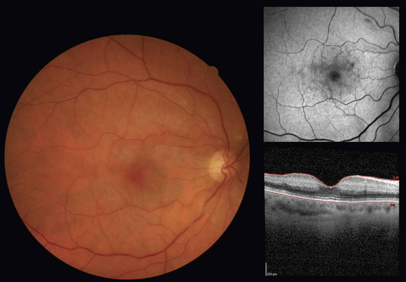
This patient presented with a VA of counting fingers OD, stating her vision had progressively worsened over the last 4 weeks and she now saw a “cloud” over her right eye. The fundus photograph showed confluent white patches throughout the macula, which are more apparent on infrared imaging (Figure 1). The OCT shows hyperreflective bands that involve the middle layers of the retina but spare the fovea, a finding known as paracentral acute middle maculopathy (PAMM).
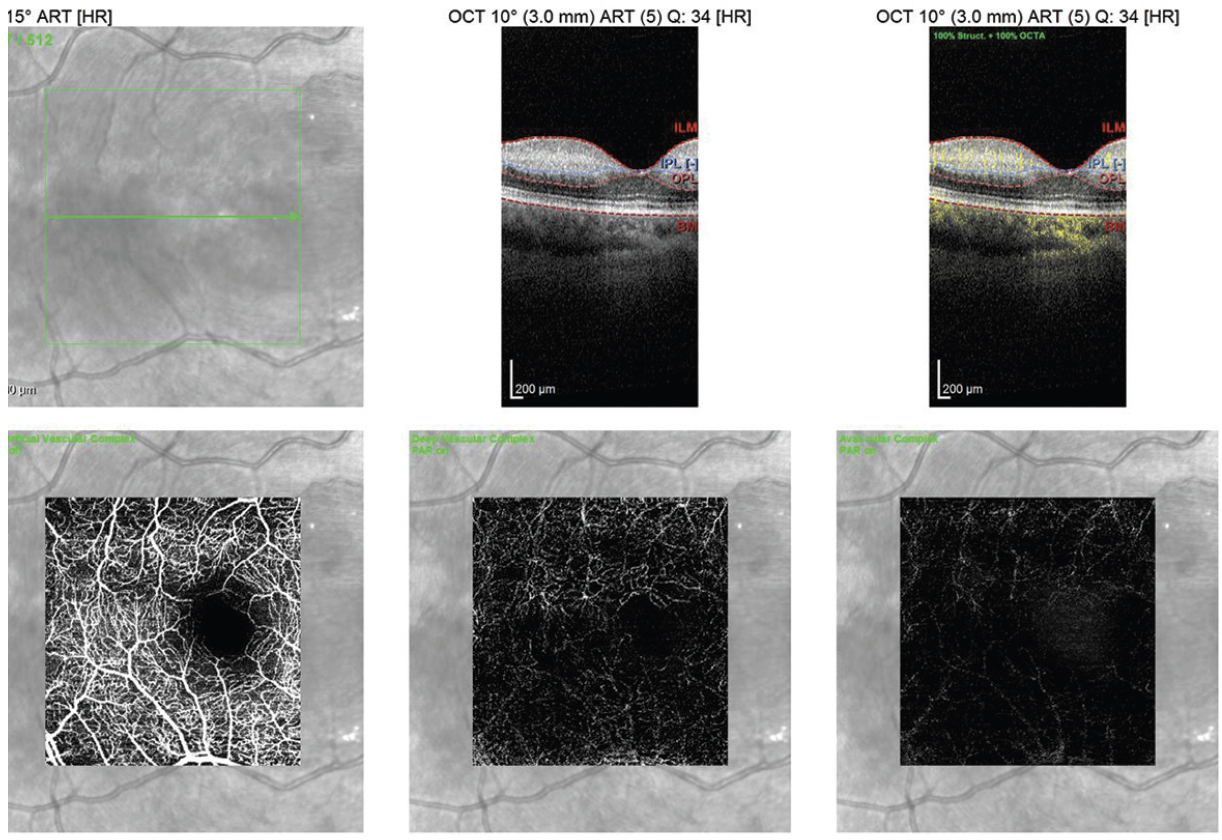
OCT angiography showed loss of capillary blood flow in the deep capillary plexus of the right eye, which is more noticeable when compared with the left eye (Figure 2). The patient was sent to her primary care provider for an urgent stroke workup, including erythrocyte sedimentation rate and C-reactive protein labs, neuroimaging, and carotid artery Doppler ultrasound, all of which were normal.
DISCUSSION
PAMM is caused by retinal ischemia specific to the deep capillary plexus. Because PAMM may be a sign of a secondary underlying condition, it is important that clinicians order an immediate systemic workup to rule out cardiovascular conditions or stroke, much like the management of a central or branch retinal artery occlusion. This may include brain MRI, carotid Dopplers, and echocardiogram. Nonetheless, PAMM may be idiopathic and can affect young healthy individuals, although the patient described here was in her 60s. Unfortunately, as with retinal arterial occlusions, there is no effective treatment for PAMM and treatment of underlying systemic disorders does not result in reversal of vision loss.


