File:Schematic representation of the hypothalamic nuclei.png
Jump to navigation
Jump to search

Size of this preview: 800 × 342 pixels. Other resolutions: 320 × 137 pixels | 640 × 274 pixels | 1,276 × 546 pixels.
Original file (1,276 × 546 pixels, file size: 539 KB, MIME type: image/png)
Captions
Captions
Add a one-line explanation of what this file represents
Summary[edit]
| Description |
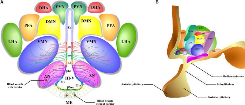
English: A schematic representation of the hypothalamic nuclei and the distribution of tanycytes over the wall of the third ventricle (III-V). (A) Coronal view of the approximate location of the hypothalamic nuclei and tanycytes. Ciliated ependymocytes (ep) line the dorsal wall of the III-V. The α1d-tanycytes (α1d) and α1v-tanycytes (α1v) have long projections that make contact with the neurons of the VMN. α2-tancycytes (α2) have projections to the AN and blood vessels. In a more ventral section of the III-V, the β1d-tanycytes (β1d) and β1v-tanycytes (β1v) make projections to the AN, making contact with orexigenic and anorexigenic neurons and blood vessels. In the floor of the III-V, the β2la-tanycytes (β2la) and β2me-tanycytes (β2me) are joined by tight junctions forming part of the median eminence (ME)-cerebrospinal fluid (CSF) barrier, and their projections make contact with the fenestrated blood vessels of the ME. (B) Sagittal view of the distribution of the hypothalamic nuclei. Ep: ependymocytes; AN: arcuate nucleus; VMN: ventromedial nucleus; DMN: dorsomedial nucleus; PVN: periventricular nucleus; DHA: dorsal hypothalamic area; PFA: perifornical area; LHA: lateral hypothalamic area; SCN: suprachiasmatic nucleus; SON: supraoptic nucleus; POA: preoptic area; MB: mammillary bodies; ME: median eminence; III-V: third ventricle. |
| Date | |
| Source | Elizondo-Vega R, Cortes-Campos C, Barahona MJ, Oyarce KA, Carril CA, García-Robles MA. The role of tanycytes in hypothalamic glucosensing. Journal of Cellular and Molecular Medicine. 2015;19(7):1471-1482. doi:10.1111/jcmm.12590. https://www.ncbi.nlm.nih.gov/pmc/articles/PMC4511346/ |
| Author | Roberto Elizondo-Vega, Christian Cortes-Campos, Maria J Barahona, Karina A Oyarce, Claudio A Carril, and Maria A García-Robles |

Licensing[edit]
This file is licensed under the Creative Commons Attribution 4.0 International license.
- You are free:
- to share – to copy, distribute and transmit the work
- to remix – to adapt the work
- Under the following conditions:
- attribution – You must give appropriate credit, provide a link to the license, and indicate if changes were made. You may do so in any reasonable manner, but not in any way that suggests the licensor endorses you or your use.
File history
Click on a date/time to view the file as it appeared at that time.
| Date/Time | Thumbnail | Dimensions | User | Comment | |
|---|---|---|---|---|---|
| current | 15:41, 15 September 2018 | | 1,276 × 546 (539 KB) | Was a bee (talk | contribs) | {{Information |Description={{en|1=A schematic representation of the hypothalamic nuclei and the distribution of tanycytes over the wall of the third ventricle (III-V). (A) Coronal view of the approximate location of the hypothalamic nuclei and tanycytes. Ciliated ependymocytes (ep) line the dorsal wall of the III-V. The α1d-tanycytes (α1d) and α1v-tanycytes (α1v) have long projections that make contact with the neurons of the VMN. α2-tancycytes (α2) have projections to the AN and blood vessel... |
You cannot overwrite this file.
File usage on Commons
The following page uses this file:
File usage on other wikis
The following other wikis use this file:
- Usage on ar.wikipedia.org
- Usage on de.wikipedia.org
- Usage on es.wikipedia.org
- Usage on fr.wikibooks.org